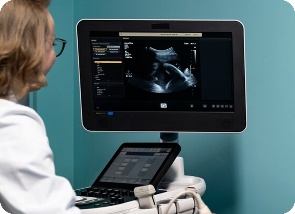
2D ECHO
Echocardiography, often referred to as 2D echocardiography or 2D ECHO, is a non-invasive imaging technique that uses ultrasound waves to create detailed images of the heart's structures and function. This method provides real-time visualizations of the heart's chambers, valves, and blood flow, allowing for the assessment of cardiac health and identification of conditions such as heart valve disease, congenital heart defects, and heart failure.
The procedure involves placing a transducer on the chest to emit and receive ultrasound waves, which are then converted into images displayed on a monitor. Advancements in 2D echocardiography technology have improved image resolution and diagnostic accuracy. 2D ECHO is a critical tool in cardiology for evaluating heart function, guiding treatment decisions, and monitoring patients with cardiovascular conditions. Its non-invasive nature and ability to provide dynamic images make it a valuable component of comprehensive cardiac care.